





How low-dose computed tomography and AI are changing lung cancer screening
As more lung cancer cases emerge among non-smokers, advances in medical imaging technology are helping doctors detect the disease earlier while reducing radiation exposure.
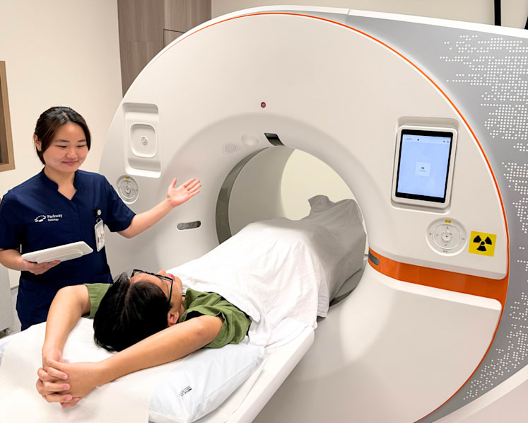
A new generation of CT scanners provides detailed images of the lungs at ultra-low radiation doses. (Photos: Parkway Radiology)

This audio is generated by an AI tool.


Presented by

Mention lung cancer and many people think of long-term heavy smoking. While smoking remains the leading risk factor, researchers have identified a concerning rise in cases among people who have never smoked, particularly older Asian women with a family history of the disease.
In Singapore, about half of all lung cancer patients have never smoked, according to a National Cancer Centre Singapore (NCCS) study. Women are disproportionately affected. Another local study also found rising rates of early-onset cases among women and an increasing incidence among female never-smokers, especially those aged 65 and above.
As the profile of lung cancer patients evolves, doctors are looking beyond smoking when assessing risk. New screening technologies, including low-dose computed tomography (CT) and artificial intelligence (AI)-powered imaging tools, are also improving early detection, helping doctors identify cancers when they are most treatable.
NOT JUST A SMOKER’S DISEASE
Data from the Singapore Cancer Registry shows that lung cancer is the third most common cancer among both men (13.2 per cent) and women (8.1 per cent). Yet more than two-thirds of cases are still diagnosed at an advanced stage.
One reason is that early-stage lung cancer often causes no symptoms. Many people are unaware they have the disease until they develop persistent chest pain, shortness of breath or start coughing up blood. By then, the cancer may have progressed to Stage 3 or 4, when survival rates are significantly lower.
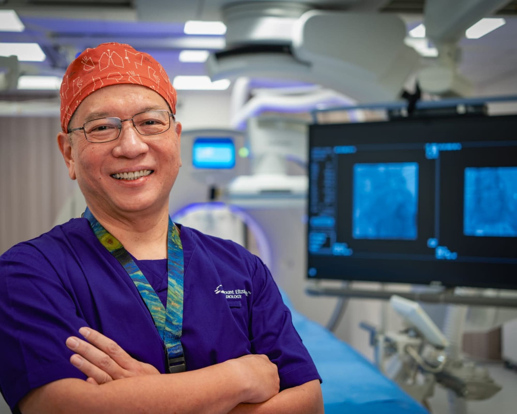
“Very early lung cancers, detected and operated on before the tumour has spread or when it has only minimally invaded the surrounding lung tissue, have an almost 100 per cent five-year survival rate,” said Dr Peter Goh, group medical director and interventional oncologist at Parkway Radiology.
In Singapore, lung cancer screening is recommended for adults aged 50 to 80 with a 20-pack-year smoking history – equivalent to smoking one pack of cigarettes a day for 20 years – who currently smoke or have quit within the past 15 years. It is currently not recommended for never-smokers.
However, emerging evidence suggests that some never-smokers may also benefit from screening. Dr Goh said middle-aged adults with additional risk factors – such as a family history of lung cancer or prolonged exposure to secondhand smoke – should discuss screening with their doctor, even if they do not smoke.
LOW-DOSE CT: ADVANCES IN LUNG CANCER SCREENING
Low-dose CT (LDCT) is widely recommended in clinical guidelines for lung cancer screening in high-risk individuals. Unlike chest X-rays, LDCT produces detailed 3D images that make small lung nodules easier to detect.
“Depending on lesion size and location, chest X-rays can miss 20 per cent to 50 per cent of early lung cancers,” said Dr Goh. “The radiation dose from LDCT is about 1mSv (millisieverts) to 2.5mSv, while a standard chest CT scan delivers about 6mSv.”
In Singapore, LDCT is being used in an ongoing lung cancer screening research study led by NCCS. As of November 2025, more than 530 participants had been screened, with Stage 1 lung cancer detected in nine individuals – all non-smokers with a family history of the disease.
HOW AI IS ASSISTING RADIOLOGISTS
Reading a CT scan is a meticulous process. Each scan produces around 400 to 700 images, and early lung cancers may appear as nodules measuring just 4mm to 8mm, said Dr Goh.
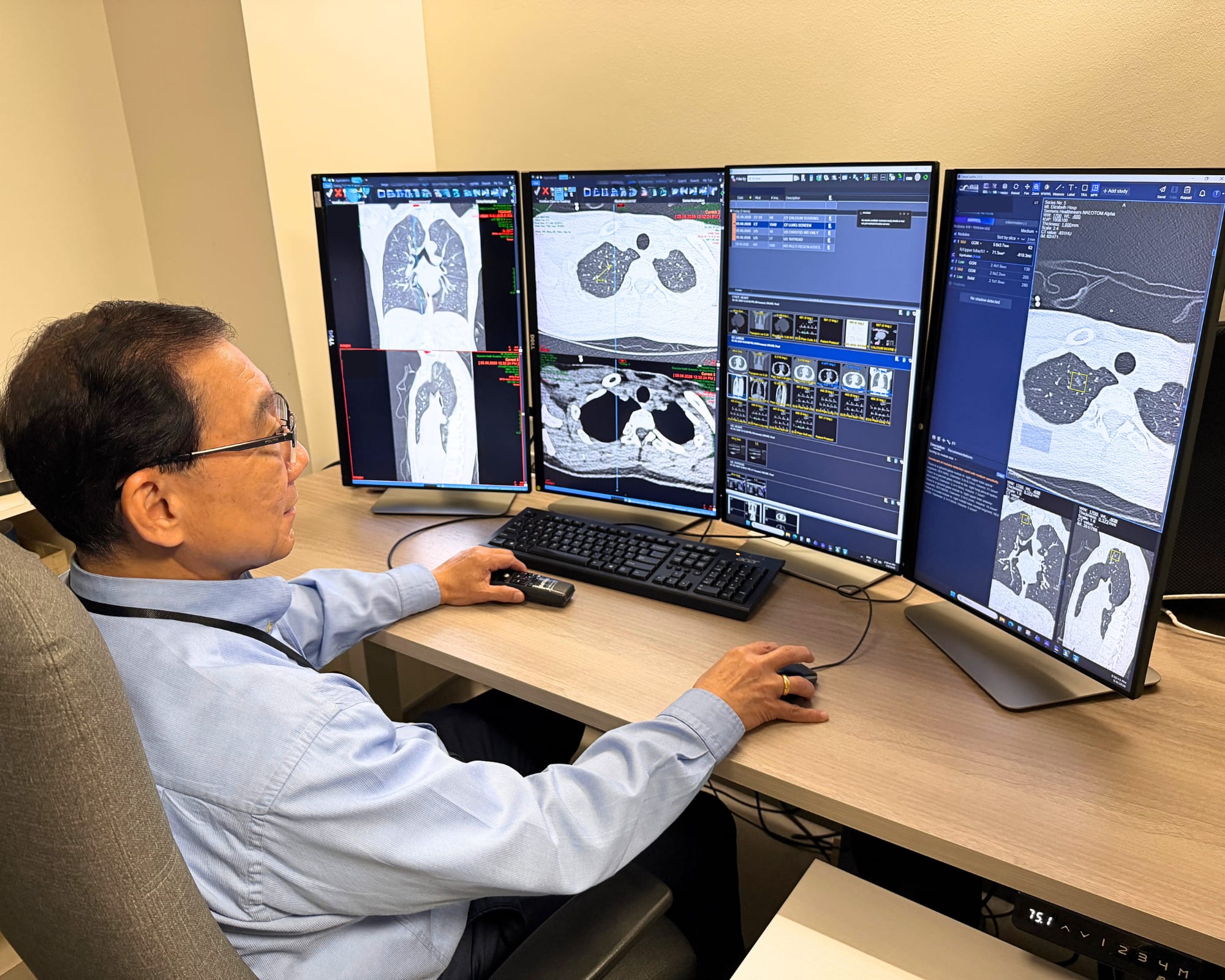
To support radiologists in analysing CT scans, Parkway Radiology uses lung CT AI software approved by the Health Services Authority across its network in IHH Healthcare Singapore hospitals. The system flags suspicious lung nodules, including those that are small or obscured, for further clinical assessment.
“AI assistance has increased our radiologists’ sensitivity for detecting early lung cancers from 76.55 per cent to 91.22 per cent,” said Dr Goh. “The AI software has been reported to improve the nodule detection rate by up to 26 per cent and reduce report generation time by up to 31 per cent.”
At Mount Elizabeth Hospital, Parkway Radiology uses photon-counting dual-source CT, a newer generation of CT imaging technology that produces high-resolution images at ultra-low radiation doses.
“Depending on the patient’s body size, the radiation dose from ultra-low-dose CT (ULDCT) can range from 0.12mSv to 0.76mSv – equivalent to one to seven conventional chest X-rays – with minimal reduction in image quality,” said Dr Goh. The scan takes less than five minutes and requires no injections or fasting.
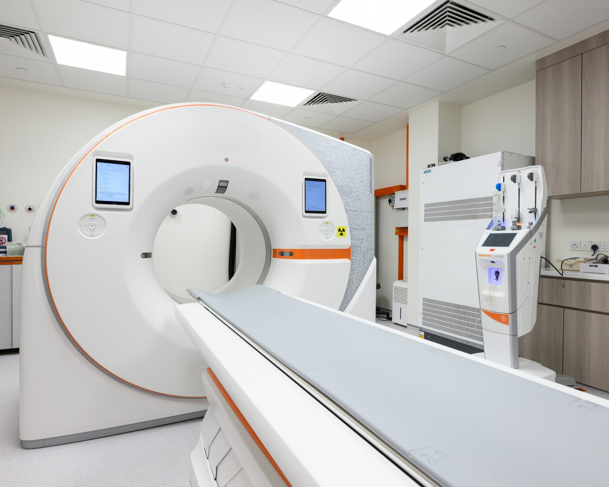
Besides Mount Elizabeth Hospital, ULDCT is also available at Gleneagles Hospital and Mount Elizabeth Novena Hospital.
EARLIER DETECTION EXPANDS TREATMENT OPTIONS
Detecting lung cancer early can broaden treatment options. When diagnosed before the disease has spread, some patients may be suitable for minimally invasive approaches that offer faster recovery than conventional surgery.
One option is interventional radiology, which uses imaging guidance to target abnormalities with greater precision. Parkway Radiology offers a range of interventional radiology services for diagnosing and treating lung conditions. These include needle biopsy to remove a small tissue sample for laboratory analysis, tumour ablation that uses extreme heat or cold to destroy cancer cells, and preoperative localisation or marking of lung nodules.
For Dr Goh, these advances all point to the same goal – finding lung cancer before symptoms develop, when treatment is most effective. “Almost every cancer we have detected early that was subsequently treated, whether through surgery or ablation, underscores the critical role of early detection in making a meaningful difference to patient outcomes,” he said.
Screening recommendations should be based on individual risk factors and made in consultation with a healthcare professional. Speak to a doctor to find out whether LDCT lung cancer screening is appropriate for you. For more information about Parkway Radiology’s services, visit parkwayradiology.com.sg, call 6388 4333 or WhatsApp 8798 8008.